Hemangiomas
Whether you are noticing a bright “strawberry” birthmark on your newborn or the sudden appearance of small, ruby-red spots on your own skin, vascular growths can be a source of confusion.
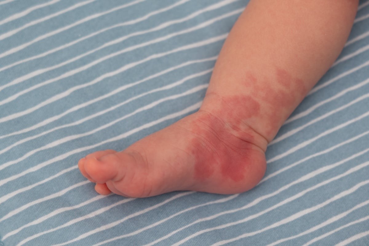
1 in 10 infants
will develop a hemangioma within the first few weeks of life
75% of individuals over age 70
exhibit cherry hemangiomas
3 to 5 times
more likely to occur in female infant than males
25%
of premature babies weighing under 1kg developing a lesion
UNDERSTANDING HEMANGIOMAS
Precision Care for Hemangiomas: Beyond the Surface of Vascular Growth
For parents and patients in Birmingham, the sudden appearance of a bright red “strawberry” mark or a deep purple vascular lesion can be a source of significant concern. Whether it is an infantile hemangioma appearing shortly after birth or a cherry hemangioma developing in adulthood, these growths are more than cosmetic “spots”—they are complex vascular proliferations.
The Anatomy of a Hemangioma
Clinically, a hemangioma is a benign (non-cancerous) tumor made up of an abnormal buildup of blood vessels. This condition is a disorder of Vascular Endothelial Proliferation. In the case of Infantile Hemangiomas, the growth is characterized by a rapid “proliferation phase” where blood vessels multiply quickly, followed by a slow “involution phase.” These lesions can be Superficial (bright red and textured), Deep (bluish or skin-colored), or Mixed, depending on which layer of the cutaneous architecture is involved.
Targeted Vascular Protocols at Inverness Dermatology
We categorize Hemangiomas (ICD-10: D18.01) by their growth pattern and depth to ensure the most effective medical intervention.
Our team focuses on Safe, Proactive Management. Treatment plans are dynamic, structured to arrest the growth of proliferating lesions and reduce the appearance of permanent vascular staining. A critical pillar of our trust promise is Functional Protection. We prioritize early intervention for lesions near sensitive areas—such as the eyes or mouth—to prevent vision or feeding obstructions, protecting the integrity of your skin and overall health.
CLINICAL DETAILS
A Breakdown of Hemangiomas
Cherry Hemangioma
Common, benign vascular proliferation in adults; also known as senile angioma.
Infantile Hemangioma
Benign vascular tumor of childhood; exhibits rapid proliferation followed by slow involution.
Port-Wine Stain (Capillary Malformation)
A congenital capillary malformation (Birthmark); does not regress and grows proportionally with the patient.
Pyogenic Granuloma
A reactive vascular lesion often following minor trauma; characterized by rapid growth and easy bleeding.
Venous Malformation
A low-flow vascular malformation present at birth; composed of abnormally formed, dilated veins.
Spider Angioma
A central red arteriole with radiating “legs” of smaller vessels; often associated with sun damage or hormonal changes.
Lentigo Maligna
(Crucial Differential) Melanoma in situ; included to emphasize the need for board-certified screening of any pigmented lesion.
EVIDENCE-BASED CARE
Our Treatment Approach
Board-certified dermatologists are specially trained to diagnose and provide customized treatments.
Hemangiomas FAQ
Clinical answers from our board-certified dermatologists.
Will my child’s hemangioma go away on its own?
Are cherry hemangiomas dangerous?
Does laser treatment for hemangiomas hurt?
Why did I suddenly develop so many cherry hemangiomas?
Accepted insurance providers